Biomolecular design through latent reasoning with RFD4-Proteina
Nathaniel Corley1,2,3*, Kieran Didi1,4,5*✉, Maxwell W. Kazman1,2,6*, Rafael I. Brent1,2,7*
Magnus S. Bauer1,2‡, Riti Biswas1,2,7‡, Julia E. Bonzanini1,2,3‡, Adam M. Chazin-Gray1,2‡, Jie Chen1,2,10‡, Maximilian Gantz1‡, Henry A. Jones11,12‡, Margaret C. Lunn-Halbert1,2,6‡, Nora K. McNamara-Bordewick1,2‡, Saman Salike1,2,19‡, DéJenaé L. See1,2,3‡, Shajesh Sharma1,2,3‡, Eric Sun1,15‡, Preetham Venkatesh1,2‡, Seth M. Woodbury1,2,3‡, Kejia Wu1,2‡, Yuyang Zhang1,8,9‡, Joel D. Allen13, Jasper Butcher1,2,10, Lemuel Chang1,2, Yehlin Cho4, Alexis Courbet1,2,15, Brian Coventry1,2, Christian Dallago4,17, Elliot S. Delfosse1,2,6, Helen E. Eisenach1,2, Andrew Favor1,2,7, Mariana Garcia Sanchez1,2, Tomas Geffner4, Inna Goreshnik1,2,15, Per Greisen18, David E. Kim1,2,15, Donghyo Kim1,2, Paul Kim1,2,7, Brianne R. King1,2, Rohith Krishna1,2, Yanjing Li1,2,10, Zi Ming Li1,3, Aiko Muraishi1,2,10, Edin Muratspahić1,2, Oliver Nagl1,16, Griffin H. Rangel1,14, Antonia Ruder1,16, Hojeong Shin1,2, Zefei Xu11,12, Yuwei Yang4, Max Crispin13, Neil P. King1,2, Frank DiMaio1,2, Gaurav Bhardwaj1,14,22, Michael Bronstein5,20, Karsten Kreis4, Donald Hilvert21, Anthony P. Green11,12, Arash Vahdat4, David Baker1,2,15✉.
Show 22 affiliations
Institute for Protein Design, University of Washington, Seattle, WA, USA
Department of Biochemistry, University of Washington, Seattle, WA, USA
Department of Bioengineering, University of Washington, Seattle, WA, USA
NVIDIA Corporation, Santa Clara, CA, USA
Department of Computer Science, University of Oxford, Oxford, UK
Graduate Program in Biological Physics, Structure and Design, University of Washington, Seattle, WA, USA
Graduate Program in Molecular Engineering, University of Washington, Seattle, WA, USA
MOE Key Laboratory of Bioinformatics, School of Life Sciences, Tsinghua University, Beijing, China
Beijing Frontier Research Center for Biological Structure, Tsinghua University, Beijing, China
Paul G. Allen School of Computer Science and Engineering, University of Washington, Seattle, WA, USA
Manchester Institute of Biotechnology, The University of Manchester, Manchester, UK
Department of Chemistry, The University of Manchester, Manchester, UK
School of Biological Sciences, University of Southampton, Southampton, UK
Department of Medicinal Chemistry, University of Washington, Seattle, WA, USA
Howard Hughes Medical Institute, University of Washington, Seattle, WA, USA
Department of Biosystems Science and Engineering, ETH Zurich, Basel, Switzerland
Department of Biostatistics and Bioinformatics, Duke University, Durham, NC, USA
BioMap Research, Palo Alto, CA, USA
Department of Chemical Engineering, University of Washington, Seattle, WA, USA
AITHYRA, Research Institute for Biomedical Artificial Intelligence, Austrian Academy of Sciences, Vienna, Austria
Laboratory of Organic Chemistry, ETH Zurich, Zurich, Switzerland
Institute for Innovations in Drug Disposition and Delivery, University of Washington, Seattle, WA, USA
* Equal contribution as co-first authors; order randomized on every refresh‡ Equal contribution as co-second authors; alphabetical by surname✉ Correspondence: Kieran Didi (kdidi@nvidia.com) and David Baker (dabaker@uw.edu)
We present RFD4-Proteina, a biomolecular design model that reasons in a shared chemical and conformational latent space covering standard and modified amino acids, sugars, DNA, and RNA. The model uses recurrent refinement to adaptively allocate computation and inference-time search guided by geometric and energy evaluations. Below are the model and its in silico benchmarks, followed by wet-lab validation.
Model Highlights
One latent space for standard and modified amino acids, sugars, DNA, and RNA, trained against force-field energies for physically plausible conformations.
Biomolecular design requires finding chemically and geometrically compatible arrangements of interacting atoms and molecules. The complexity of problems which can be tackled by deep learning design models is limited by both the molecular landscape encompassed by the model and the ability to explore that landscape effectively. Here we introduce RFD4-Proteina, which addresses these limitations through intrinsic and extrinsic reasoning in a shared, physics-informed chemical and conformational latent space. The latent space encompasses standard and modified amino acids, sugars, DNA, and RNA, supervised with force-field energies to favor physically plausible conformations. Within this space, intrinsic reasoning uses recurrent refinement to adaptively allocate computation, while extrinsic reasoning guides search with arbitrary constraints such as energetics or geometric criteria. Across in silico protein design benchmarks, RFD4-Proteina achieves higher design success rates at greater generation throughput than all methods evaluated, with further gains from conformational flexibility, recurrent refinement, and search. Without additional training, the same model performs equivalently to or better than state-of-the-art specialist methods for related tasks, including side-chain packing, fixed-backbone sequence design, conformational sampling, and design refinement. Experimental validation spans specific and cross-reactive binding to therapeutically relevant targets, modified and glycosylated proteins, and catalysis of both bond-breaking and bond-forming reactions, including a first proof-of-concept demonstration of a de novo designed enzyme for active pharmaceutical ingredient synthesis. By uniting diverse molecular chemistry, conformational flexibility, scalable reasoning, and physical modeling with machine-learning generation, RFD4-Proteina expands the frontier of protein design.
Unified Latent Reasoning
RFD4-Proteina makes use of a unified latent space. Select from the tabs below to explore the latent space, a full generation trajectory, and intrinsic and extrinsic reasoning.
An autoencoder represents amino acids, sugars, and nucleotides in
a shared 32-dimensional chemical and conformational space.
60 denoising steps · 120, 180, and 300 trunk evaluations
Trunk evaluations per step
Current low, medium, and high thinking presets allocate more recurrent updates to earlier steps. Medium is the default. Recycling schedule guide.
Different molecular properties become resolved at different points
in the generation trajectory. In this example, we select
branches first by interface area, which becomes resolved early in
generation, followed by hydrogen bonding as finer-scale contacts
emerge, and finally by AlphaFold3 (AF3) confidence.
Typically, design methods trade accuracy for speed. For example, hallucination methods such as BindCraft achieve strong pass rates but only produce tens of designs per GPU-hour. Generative diffusion models, meanwhile, produce hundreds to thousands of designs per GPU-hour but exhibit lower per-design quality. On all four benchmarks, RFD4-Proteina achieves both higher pass rates and higher throughput than every other method evaluated, even at its lowest thinking setting.
For detailed results, evaluation criteria, and method comparisons, explore the full benchmark section.
What counts as a pass. We refold each designed sequence with AlphaFold3 (AF3) and check it against the intended structure and task-specific criteria. The clustered pass rate counts distinct folds only, so repeating one success earns nothing.
Better and faster. For protein binding, high-thinking, single-shot RFD4-Proteina achieves a 10.6% clustered pass rate versus 5.9% for the next-best model (BindCraft), at 42-fold higher throughput. For small molecules it is 6.1-fold higher than the next-best model (14.3% versus 2.4%). For enzyme scaffolding it reaches 12.1% versus 5.7%, and for DNA binding 3.5% versus 0.8% for BoltzGen and 0.5% for fixed-DNA RFD3.
Solves more design targets. RFD4-Proteina produces passing designs for more targets than any individual baseline across all four benchmarks: 22 of 24 protein targets, all 77 small-molecule targets, 35 of 37 enzyme tasks, and all three DNA targets (versus two for fixed-DNA RFD3). For small molecules, the next-best model, RFD3, solves 64 of 77 targets.
Thinking longer helps. From low to high thinking, the protein-binding clustered pass rate rises from 7.7% to 10.6%, while throughput decreases from 2,388 to 986 designs per GPU-hour.
Figure 2. Clustered pass rate against generation throughput (designs per GPU-hour) for protein binding (24 targets), small-molecule binding (77), enzyme active-site scaffolding (37), and DNA binding (3). Right-hand bars show target coverage and passing clusters per GPU-hour including AF3 evaluation. Low, medium, and high thinking denote task-specific recurrent-refinement budgets without structure-predictor feedback.
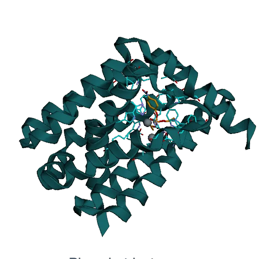
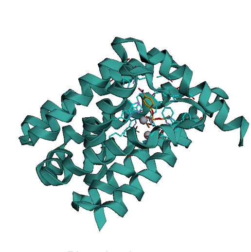
The structures below are computational models of designs we tested in the lab. Drag to rotate, scroll to zoom, and use the buttons to switch designs or states.
Calmodulin: calcium-dependent binders
Calmodulin switches conformation when it binds Ca2+, and many of its partners recognize only one state. We asked RFD4-Proteina for binders to the calcium-bound form only.
Hit rate. 12 of 18 designs bound with an apparent KD below 1 µM and showed no detectable binding without calcium.
Affinity. Apparent KD from 69 pM* to 37.0 nM. The interactive example above binds at 2.48 nM.
Figure 4a. Calcium-dependent calmodulin binding: the design concept, the distribution of apparent KD across designs, and the paper’s 69 pM example* with BLI responses in Ca2+ (teal) and EDTA (gray). The interactive viewer above shows a different binder. Full figure
* The paper’s 69 pM apparent affinity comes from a 1:1 fit at the off-rate fitting floor; its precise affinity and ranking remain unresolved.
Peptide–MHC binders
A binder to a peptide–MHC complex must recognize a few exposed peptide residues while ignoring the conserved MHC around them. Cancerous and infected cells are flagged this way, which is why look-alike peptides from healthy cells are the main risk.
Targets. MART-1, SARS-CoV-1 membrane-protein, and Phox2B peptides.
Affinity. Yeast-display titrations with pMHC tetramers gave apparent half-maximal binding concentrations of 340 pM, 710 pM, and 1.60 nM.
Specificity. Over 100-fold preference for the on-target peptide in all cases. The Phox2B and SARS-CoV-1 off-targets differ from the target peptide by only one and two residues.
Figure 4b. Binders to MART-1, SARS-CoV-1, and Phox2B peptide–MHC complexes: design models, yeast-display titrations, and binding to the six peptide–MHC targets in the specificity screen. Full figure
Disordered peptide binders
Intrinsically disordered regions are involved in neurodegeneration, chronic pain, and cancer, but they have no fixed structure to design against. We targeted eight disordered peptides: five known to fold when bound and three without that tendency.
Hit rate. Tight binders for four of five peptides known to adopt secondary structure when bound, and two of three peptides without that tendency.
First binders. To our knowledge, the first de novo binders to the tested YAP1 and Tau epitopes. The strongest hits have SPR affinities of 112 pM and 527 pM.
Specificity. All-by-all tests of the best hits against five targets showed a preference for the intended peptide in every case, with varying strength.
Figure 4c,d. Binders to four disordered peptides (YAP1, Tau153, N17, PER2) with SPR sensorgrams and 1:1 fits (c), and all-by-all SPR specificity of the best binders against five peptides (d). Full figure
TrkA: species and paralogue specificity
Trk receptors are targets in neuropathic pain and in cancers such as neuroblastoma. Human TrkA is an established benchmark, so we asked for more: binders that avoid mouse TrkA (86% sequence identity over the modeled region) and the human paralogues TrkB and TrkC.
Human-specific. The four strongest hTrkA binders showed no binding to any off-target.
Cross-reactive. 18 of 94 designs bound human and mouse TrkA but not TrkB or TrkC. The best was sub-nanomolar for hTrkA and 37.7 nM for mTrkA.
Unconstrained hTrkA. 158 of 188 designs (84%) were sub-micromolar, against 18 of 60 for the next most successful campaign.
TrkB and TrkC. One sub-micromolar TrkB binder (30.2 nM) and 20 TrkC screening hits. To our knowledge, the first designed binders to either.
Figure 4e,f. Human-specific and human/mouse cross-reactive TrkA campaigns with SPR and BLI measurements against human and mouse TrkA (e), and the experimental hit rate of unconstrained human TrkA design compared with other campaigns (f). Full figure
Glycan shielding
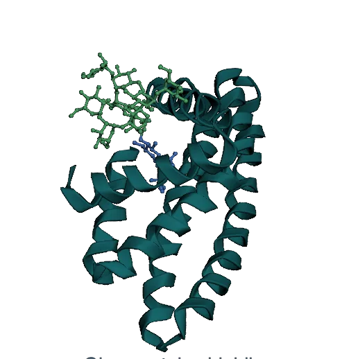
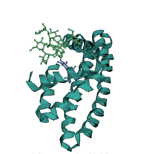
N-linked glycans are trimmed by enzymes in the ER and Golgi. Blocking that processing triggers an immune response to the underprocessed glycan, which makes it usable as a vaccine adjuvant. A design has to shield the core GlcNAc while keeping the terminal mannose accessible.
Previous work.Lunn-Halbert et al. designed proteins that retained Man5–Man8 glycans; none fully blocked processing to Man9 or Glc1Man9.
In silico. Higher design success rates than the earlier RFDAA-based method.
Plate-scale screen. Glycoproteins retained mostly Man9 and Glc1Man9. 8 of 9 designs had more than 90% oligomannose on occupied sites, with smaller proteins than before.
Figure 5a–c. Shielding the core of an N-linked glycan while exposing the terminal mannose (a), design B10 with its glycan composition (b), and the extent of underprocessing across design campaigns (c). Full figure
Phosphopeptide binders
Phosphorylation regulates molecular interactions across all domains of life. Bauer et al. demonstrated the design of phosphorylation-dependent binders to phosphotyrosine-containing peptides. Here, we targeted three phosphorylated variants of the histone H3 tail: pS10, pT11, and pS28.
Binders. Phosphorylation-dependent binders for all three sites.
Selectivity. The pT11 and pS10 hits preferred their phosphorylated targets and stayed selective against phosphorylation at the adjacent site.
Titrations. Apparent response midpoints of 179 nM (pS10) and 133 nM (pT11) in yeast display. These are response midpoints, rather than equilibrium affinities.
Figure 5d–f. Binders to histone H3 pS10 (d) and pT11 (e) with yeast-display titrations of the phosphorylated and unmodified peptides, and selectivity between adjacent phosphorylation sites by NGS recovery (f). Full figure
Phosphotriesterases
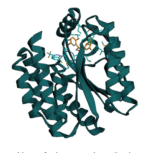
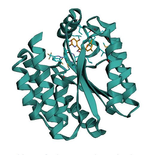
Paraoxon is an organophosphate pesticide and a potent neurotoxin. RFD3 demonstrated the design of active phosphotriesterases by scaffolding six catalytic residues around two metal ions, achieving kcat/KM up to 4 ± 3 M−1 s−1. Building on this work, we used RFD4-Proteina to scaffold an expanded, eight-residue active site, testing whether additional catalytic interactions could improve activity.
In silico. For the eight-residue active site, 111 of 1,000 RFD4-Proteina designs passed the computational filters, compared with 1 of 10,000 for RFD3.
Screening. 89 of 288 RFD4-Proteina designs were active, spanning 25 of 47 scaffold families.
Best design. Of six characterized designs, the best reached kcat/KM = 3,600 ± 500 M−1 s−1 and kcat = 0.9 s−1, a 1.5 × 10⁷-fold rate enhancement over uncatalyzed hydrolysis. The six characterized designs showed 10- to 1,000-fold higher catalytic efficiencies than the earlier six-residue designs. The numbers on the viewer buttons are each design’s kcat/KM in M−1 s−1.
Figure 6a–d. Paraoxon hydrolysis (a), the eight-residue active-site theozyme (b), in silico success rates for RFD3 and RFD4-Proteina (c), and four designs with their backbone and motif RMSDs and catalytic efficiencies (d). Full figure
Ligase for lenacapavir synthesis
Lenacapavir is an HIV-prevention drug whose manufacturing cost limits access. One step joins an activated ester to the (S)-enantiomer of a hindered chiral amine, so the enzyme has to favor aminolysis over hydrolysis of the ester.
Design. We scaffolded a cysteine-protease catalytic triad and used multi-state AF3 filtering to bias designs toward aminolysis.
Hit rate. Four of 67 designs formed the amide above background. An earlier RFD3 campaign found none in 72.
Best design. Design 52 converted 79.0% of the (S)-amine and 8.8% of the (R)-amine, with acid at the no-enzyme level (0.29 vs 0.26 mM). Design 58 gave 61.7% vs 14.6%.
Significance. To our knowledge, the first proof of concept of a de novo enzyme for a step in active pharmaceutical ingredient synthesis.
Figure 6h–n. The lenacapavir amide-forming step (h), the ligase input motif (i), in silico success rates (j), designs 52 and 58 (k), HPLC traces (l), conversion by parent designs and catalytic-Cys knockouts (m), and conversion of the (S)- and (R)-amine (n). Full figure
Code & Weights
Run RFD4-Proteina
Code on GitHub: inference, reward-guided search, training, and evaluation.
Documentation: inputs, running, search, metrics, and the API reference.
Benchmarks for arbitrary views on the underlying data sources.
Citation
RFD4-Proteina:
@article{corley2026rfd4proteina,
  title        = {Biomolecular design through latent reasoning with {RFD4-Proteina}},
  author       = {Corley, Nathaniel and Didi, Kieran and Kazman, Maxwell W. and Brent, Rafael I. and others},
  year         = {2026},
  note         = {Preprint},
  url          = {https://rfd4-proteina.org/assets/paper/RFD4-Proteina.pdf}
}