PyPI package
pip install "rfd4-proteina[metrics-cuda13]"






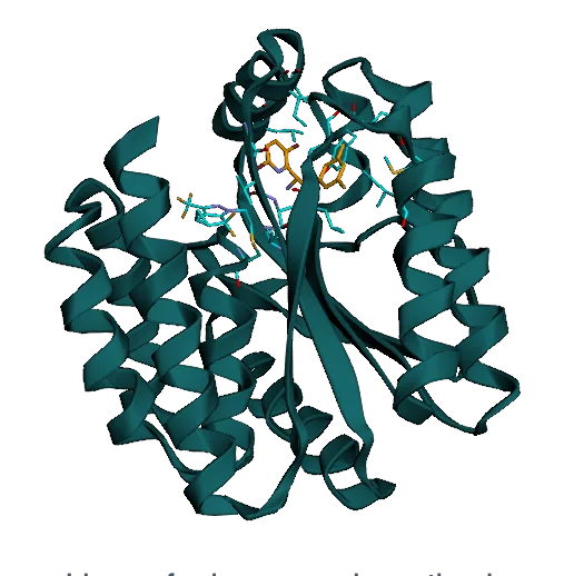
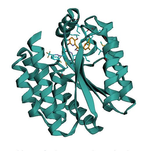
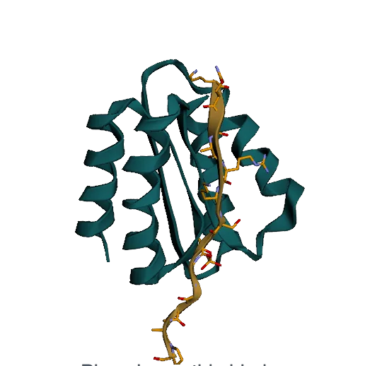
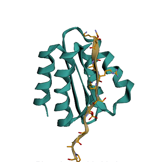
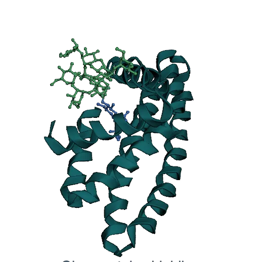
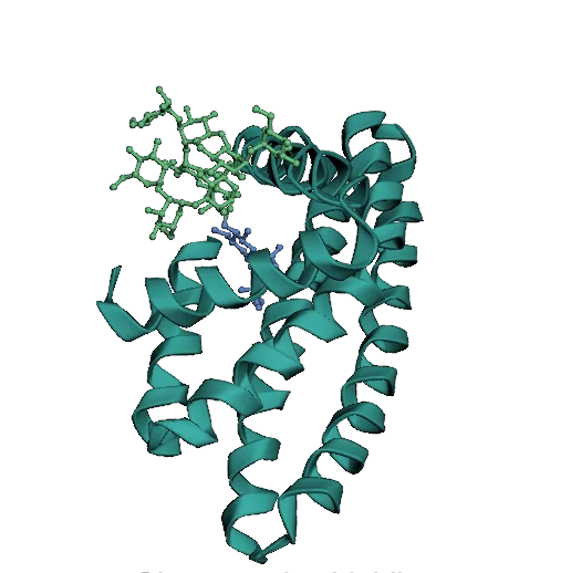
RFD4-Proteina is available as a Python package on PyPI and as a Hugging Face Diffusers pipeline. Both need Python 3.12 and an NVIDIA GPU with a CUDA 13-compatible driver; the PyPI release and the Hugging Face repositories go public with the release. The quickstart in the docs covers the pip package, a git clone with make setup, and Diffusers.
pip install "rfd4-proteina[metrics-cuda13]"
pip install diffusers \
    git+https://huggingface.co/nvidia/RFD4-Proteina-prep

import torch
from diffusers import RFD4ProteinaPipeline
from rfd4_proteina_prep import prepare_cif, write_cif

pipe = RFD4ProteinaPipeline.from_pretrained(
    "nvidia/RFD4-Proteina-Diffusers", dtype=torch.bfloat16).to("cuda:0")
features, atoms = prepare_cif("input.cif", seed=42, device=pipe.device)
output = pipe(features, generator=torch.Generator("cuda:0").manual_seed(42))
write_cif(output, features, atoms, "design.cif")
Overview. We present RFD4-Proteina, a biomolecular design model that reasons in a shared chemical and conformational latent space covering standard and modified amino acids, sugars, DNA and RNA. Recurrent refinement lets it spend more computation where denoising is hard, and search lets it satisfy arbitrary constraints. Below are the model and its in silico benchmarks, followed by wet-lab validation.
Shared Latent Space
Amino acids, sugars, DNA and RNA in one representation, supervised with force-field energies.
Latent Reasoning
Recurrent refinement (intrinsic) and constraint-guided search (extrinsic).
Enzymes
89 of 288 phosphotriesterase designs active. A designed ligase for a lenacapavir synthesis step.
Specific Binders
158 of 188 sub-micromolar hTrkA designs. 112 pM to a disordered YAP1 peptide.
Biomolecular design requires finding chemically and geometrically compatible arrangements of interacting atoms and molecules. The complexity of problems which can be tackled by deep learning design models is limited by both the molecular landscape encompassed by the model and the ability to explore that landscape effectively. Here we introduce RFD4-Proteina, which addresses these limitations through intrinsic and extrinsic reasoning in a shared, physics-informed chemical and conformational latent space. The latent space encompasses standard and modified amino acids, sugars, DNA, and RNA, supervised with force-field energies to favor physically plausible conformations. Within this space, intrinsic reasoning uses recurrent refinement to adaptively allocate computation, while extrinsic reasoning guides search with arbitrary constraints such as energetics or geometric criteria. Across in silico protein design benchmarks, RFD4-Proteina achieves higher design success rates at greater generation throughput than all methods evaluated, with further gains from conformational flexibility, recurrent refinement, and search. Without additional training, the same model performs equivalently to or better than state-of-the-art specialist methods for related tasks, including side-chain packing, fixed-backbone sequence design, conformational sampling, and design refinement. Experimental validation spans specific and cross-reactive binding to therapeutically relevant targets, modified and glycosylated proteins, and catalysis of both bond-breaking and bond-forming reactions, including a first proof-of-concept demonstration of a de novo designed enzyme for active pharmaceutical ingredient synthesis. By uniting diverse molecular chemistry, conformational flexibility, scalable reasoning, and physical modeling with machine-learning generation, RFD4-Proteina expands the frontier of protein design.
RFD4-Proteina designs inside one latent space. Pick a tab to see the space itself, a full generation trajectory, and the two kinds of reasoning that run on top of it.
An autoencoder represents amino acids, sugars, and nucleotides in a shared 32-dimensional chemical and conformational space. A decoder reconstructs their atoms, with Rosetta-energy supervision to favor physically plausible conformations.

An enzyme-design trajectory for cleavage of a peptide–DNA linkage. Circular arrows mark recurrent passes; search criteria are applied at successive stages.
Recurrent updates refine each denoising step.
60 denoising steps · 110, 185 and 295 trunk evaluations
Trunk evaluations per step
Branches are selected by interface area, then hydrogen bonding, then AF3 confidence, as these properties become resolved.

Design methods usually trade accuracy for speed. Hallucination methods such as BindCraft reach good pass rates but produce only tens of designs per GPU-hour; diffusion models produce hundreds to thousands but pass less often. On all four benchmarks, RFD4-Proteina is better on both counts: even at its lowest thinking setting, it has a higher pass rate and higher throughput than every other method we compared. In the figure below, the green region marks methods that are better on both counts.
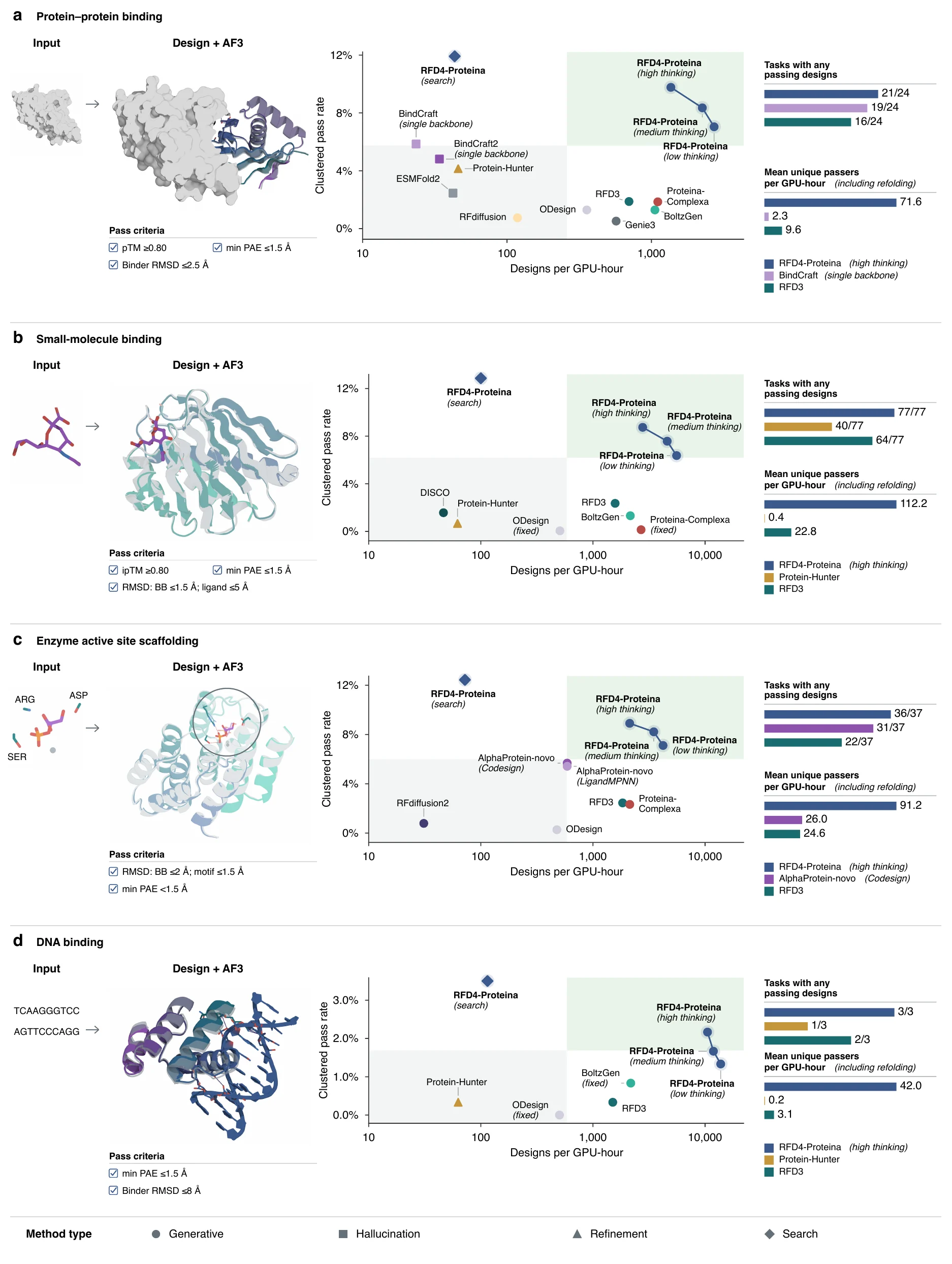
Figure 2. Clustered pass rate against generation throughput (designs per GPU-hour) for protein binding (24 targets), small-molecule binding (77), enzyme active-site scaffolding (37) and DNA binding (3). Right-hand bars show target coverage and passing clusters per GPU-hour including AF3 evaluation. Diamonds denote search guided by a structure predictor.
The structures below are computational models of designs we tested in the lab (designs, refolded structures or Protenix predictions), not experimental structures. Drag to rotate, scroll to zoom, and use the buttons to switch designs or states.
Calmodulin switches conformation when it binds Ca2+, and many of its partners recognize only one state. We asked RFD4-Proteina for binders to the calcium-bound form only.
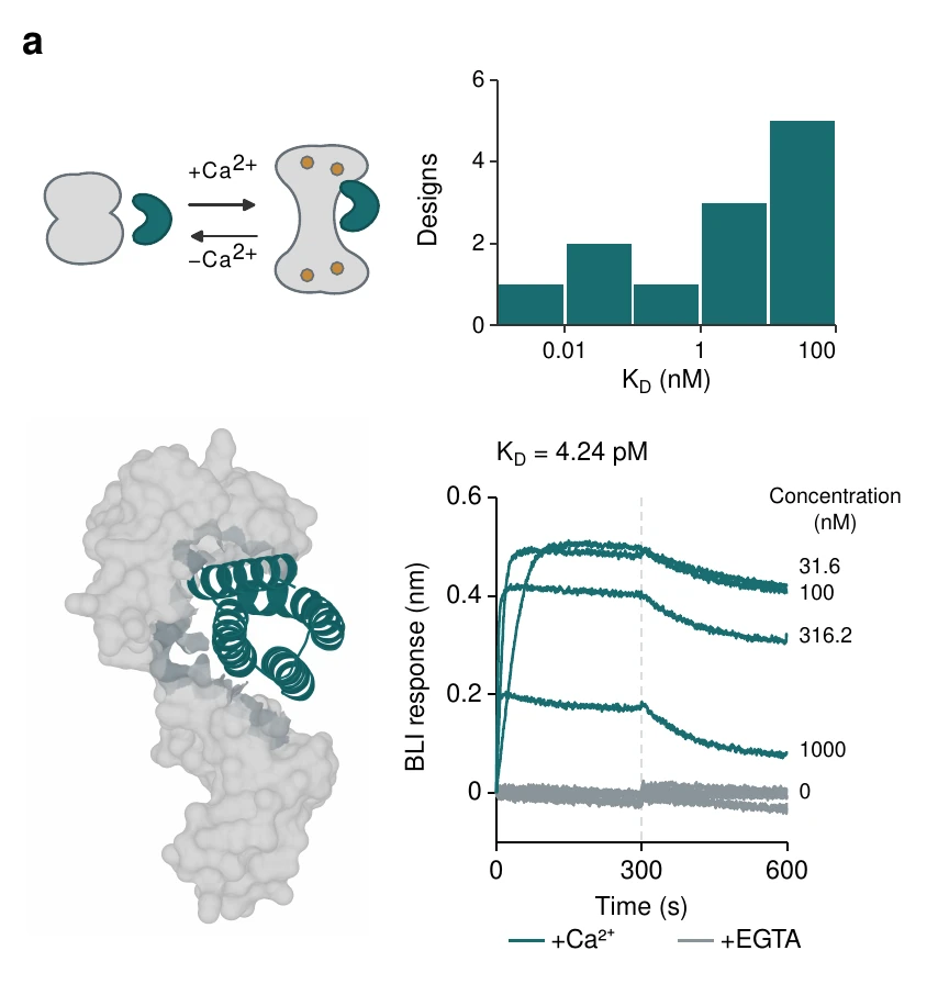
Figure 4a. Calcium-dependent calmodulin binding: the design concept, the distribution of apparent KD across designs, a designed binder on calmodulin, and the BLI response of the strongest binder with Ca2+ (teal) and with EGTA (gray). Full figure
A binder to a peptide–MHC complex has to read a few exposed peptide residues and ignore the conserved MHC around them. Cancerous and infected cells are flagged this way, which is why look-alike peptides from healthy cells are the main risk.
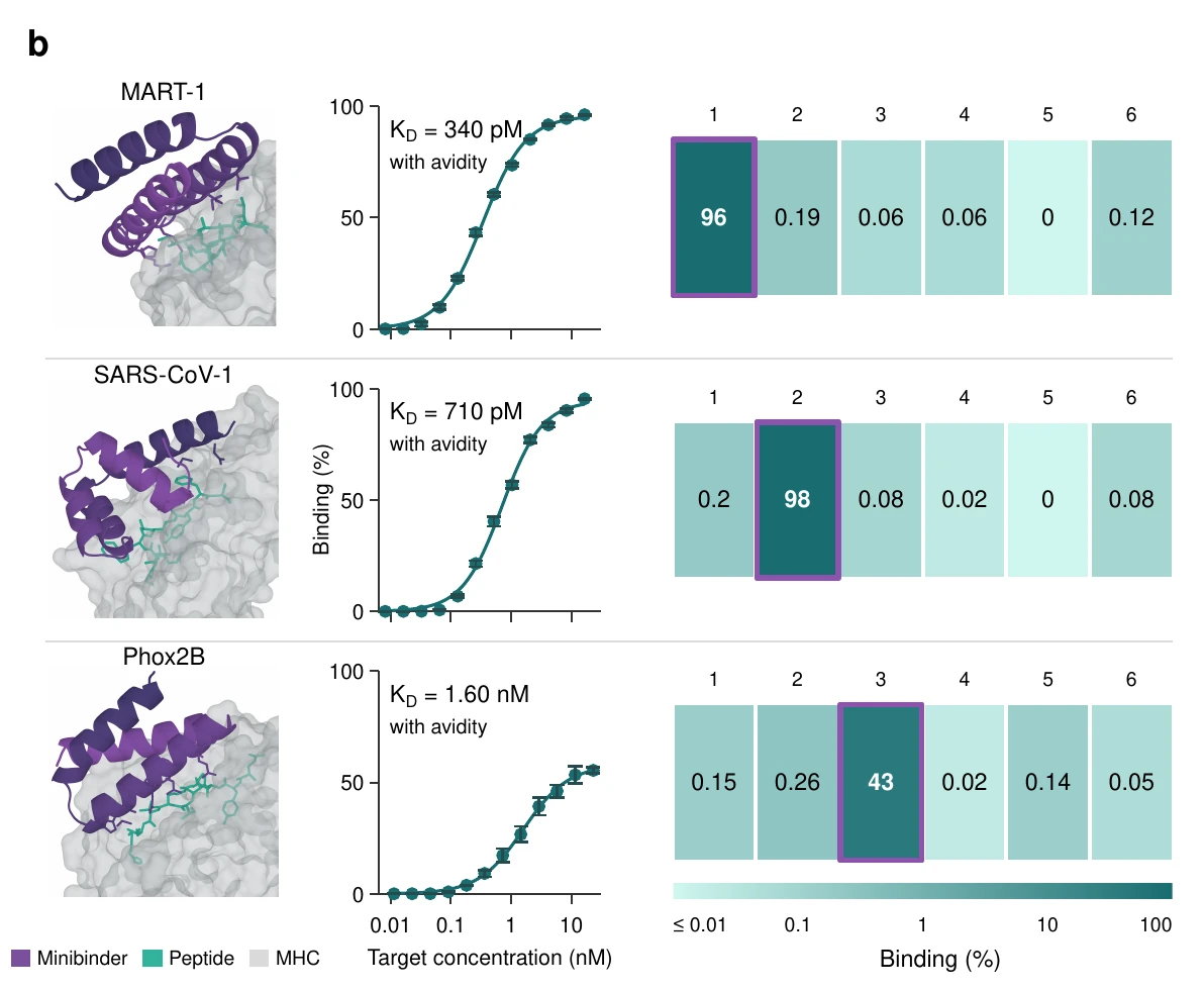
Figure 4b. Binders to MART-1, SARS-CoV-1 and Phox2B peptide–MHC complexes: design models, yeast-display titrations, and binding to the six peptide–MHC targets in the specificity screen. Full figure
Intrinsically disordered regions are involved in neurodegeneration, chronic pain and cancer, but they have no fixed structure to design against. We targeted eight disordered peptides: five known to fold when bound and three without that tendency.
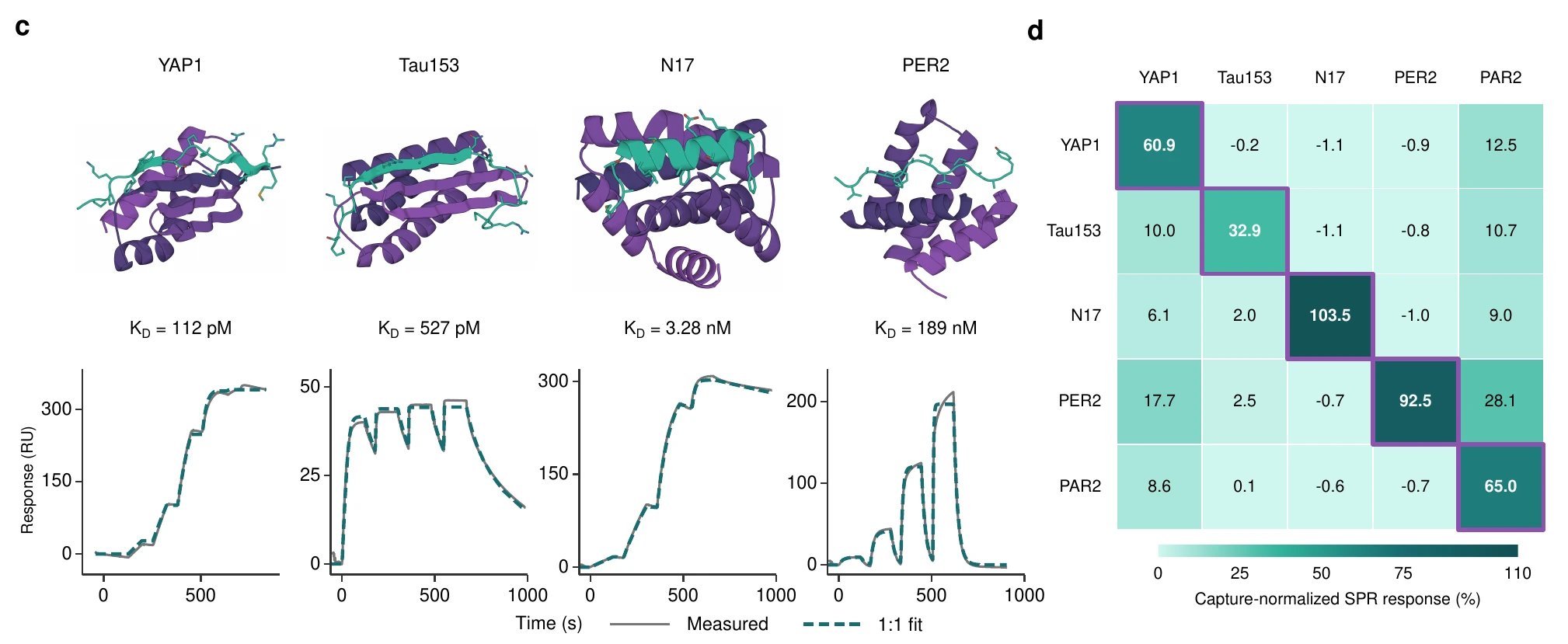
Figure 4c,d. Binders to four disordered peptides (YAP1, Tau153, N17, PER2) with SPR sensorgrams and 1:1 fits (c), and all-by-all SPR specificity of the best binders against five peptides (d). Full figure
Trk receptors are targets in neuropathic pain and in cancers such as neuroblastoma. Human TrkA is an established benchmark, so we asked for more: binders that avoid mouse TrkA (86% sequence identity over the modeled region) and the human paralogues TrkB and TrkC.
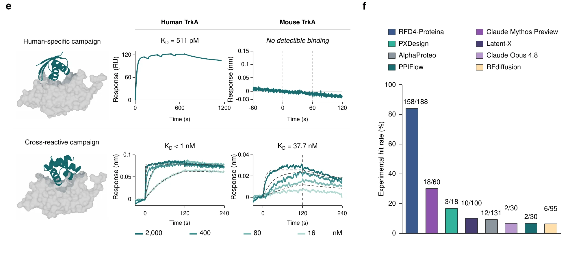
Figure 4e,f. Human-specific and human/mouse cross-reactive TrkA campaigns with BLI against human and mouse TrkA (e), and the experimental hit rate of unconstrained human TrkA design compared with other campaigns (f). Full figure
N-linked glycans are trimmed by enzymes in the ER and Golgi. Blocking that processing triggers an immune response to the underprocessed glycan, which makes it usable as a vaccine adjuvant. A design has to shield the core GlcNAc while keeping the terminal mannose accessible.
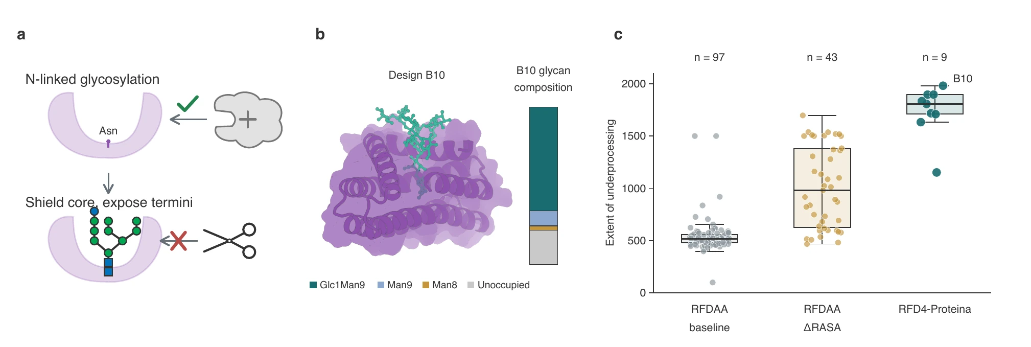
Figure 5a–c. Shielding the core of an N-linked glycan while exposing the terminal mannose (a), design B10 with its glycan composition (b), and the extent of underprocessing across design campaigns (c). Full figure
Phosphorylation regulates molecular interactions across all domains of life. We targeted three phosphorylated variants of the histone H3 tail: pS10, pT11 and pS28.
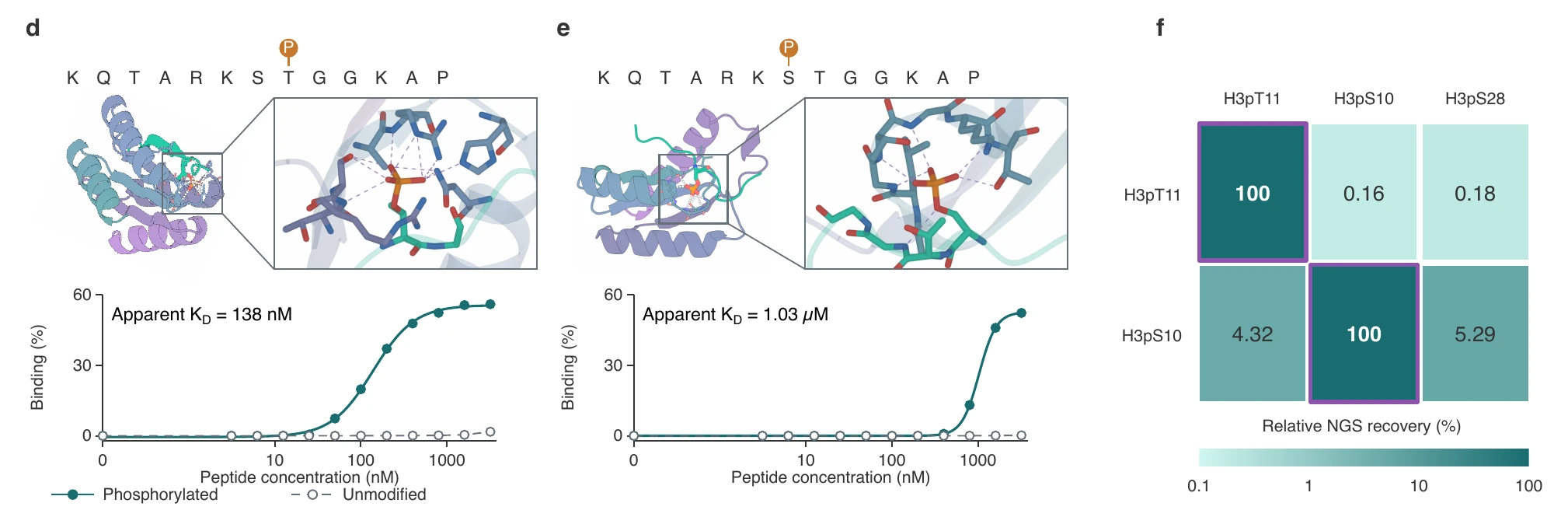
Figure 5d–f. Binders to histone H3 pT11 (d) and pS10 (e) with yeast-display titrations of the phosphorylated and unmodified peptides, and selectivity between adjacent phosphorylation sites by NGS recovery (f). Full figure
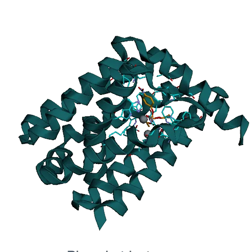
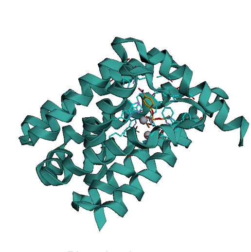
Paraoxon is an organophosphate pesticide and a potent neurotoxin. Our earlier RFD3 designs scaffolded six catalytic residues around two metal ions and reached kcat/KM of only 4 ± 3 M−1 s−1. We added two catalytic residues, which makes the active site harder to scaffold.
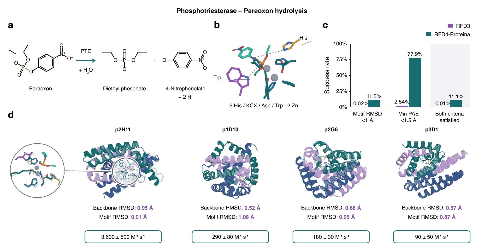
Figure 6a–d. Paraoxon hydrolysis (a), the eight-residue active-site theozyme (b), in silico success rates for RFD3 and RFD4-Proteina (c), and four designs with their backbone and motif RMSDs and catalytic efficiencies (d). Full figure
Lenacapavir is an HIV-prevention drug whose manufacturing cost limits access. One step joins an activated ester to the (S)-enantiomer of a hindered chiral amine, so the enzyme has to favor aminolysis over hydrolysis of the ester.
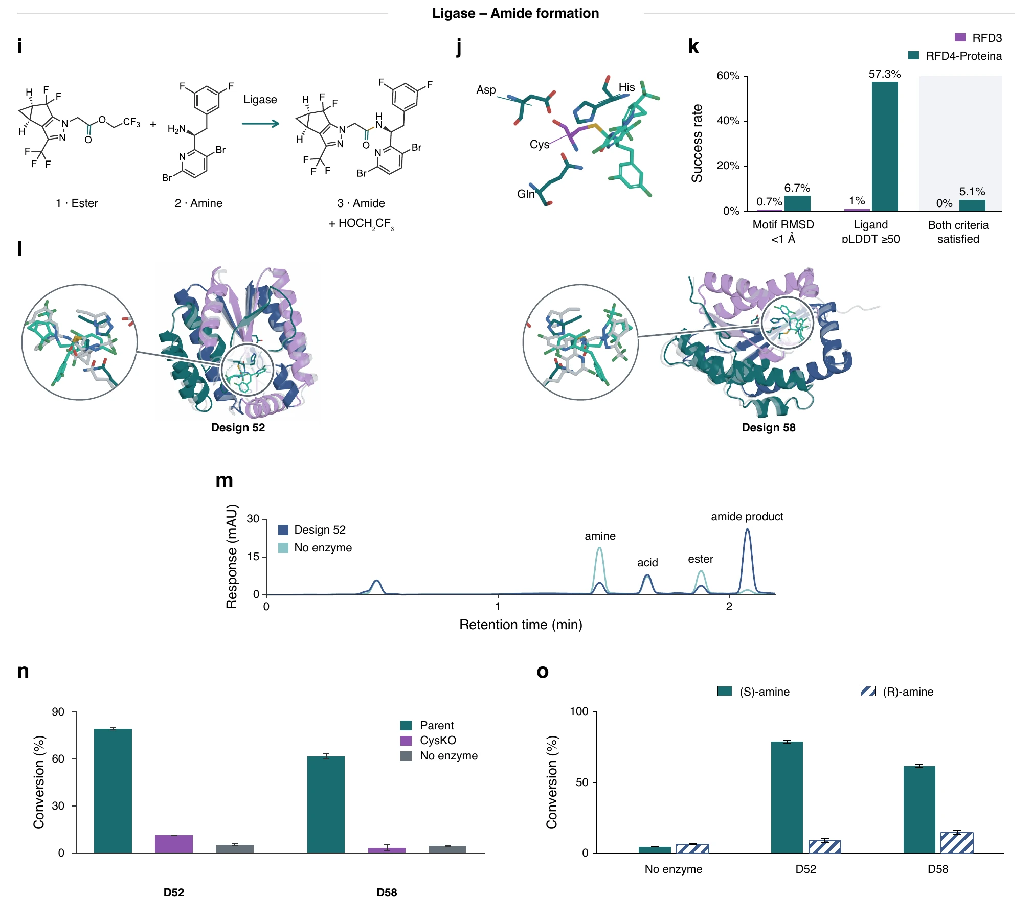
Figure 6i–o. The lenacapavir amide-forming step (i), the cysteine-protease theozyme (j), in silico success rates (k), designs 52 and 58 (l), HPLC traces (m), and conversion of the (S)- and (R)-amine (n,o). Full figure
@article{corley2026rfd4proteina,
  title        = {Biomolecular design through latent reasoning with {RFD4-Proteina}},
  author       = {Corley, Nathaniel and Didi, Kieran and Kazman, Maxwell W. and Brent, Rafael I. and others},
  year         = {2026},
  note         = {Preprint forthcoming}
}
